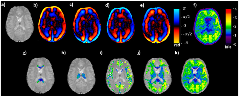
Figure 2:

Magnitude image of a axial slice (a) demonstrating snapshot of wave images at four time points in (b) through (e), the corresponding isotropic stiffness map in (f) and isotropic stiffness map excluding the lateral ventricles in each ROI of corpus callosum in (g), thalamus in (h), gray matter in (i), white matter in (j) and whole brain in (k).
