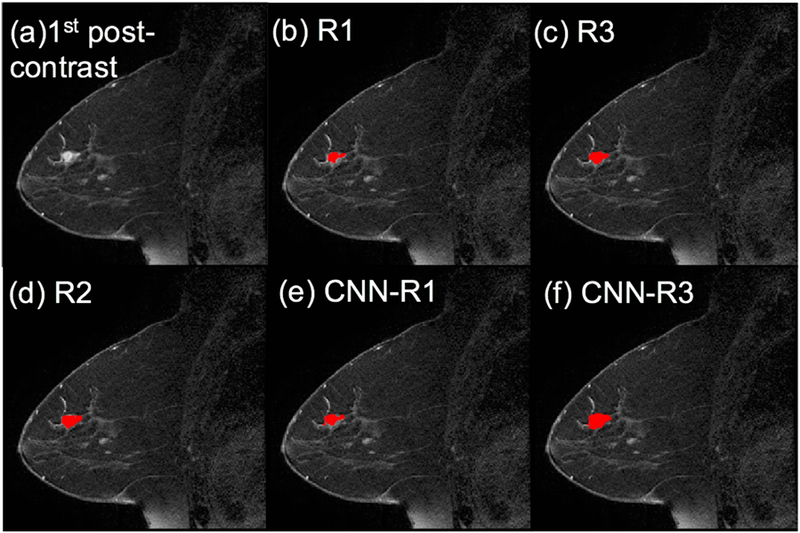
Figure 2.

ROI comparison of an example from the testing set. (a) first post-contrast frame, (b)~(f) lesion segmentations are superimposed on the first post-contrast frame in red (the manual ROIs from the three radiologists and the CNN-based ROIs trained by R1’s and R3’s segmentations, respectively).
