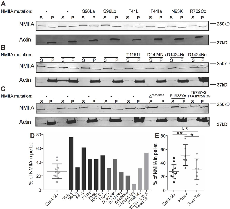
Figure 4.

Association of NMIIA with the RBC membrane skeleton in MYH9-RD patients. (A-C) RBC Mg++ ghosts from normal donor shipped controls and MYH9-RD motor (A), coiled-coil rod (B), and non-helical tail (C) domain patients were extracted in Triton X-100 buffer followed by SDS-PAGE and immunoblotting for NMIIA heavy chain (top) and actin (bottom) in the supernatant (S) and Triton-insoluble membrane skeleton pellet (P) fractions. Each pair of lanes represents a unique normal donor or patient, in the same order as in Figure S9. (D) Quantification of % NMIIA in the membrane skeleton pellet fraction, comparing normal shipped controls (dot plot) with individual motor domain patients (black bars), coiled-coil rod domain patients (dark gray bars), and non-helical tail domain patients (light gray bars). Each dot in the dot plot and each bar in the bar graph represents one technical replicate of each sample. (E) Quantification in (D) grouped by normal donor shipped controls, motor domain patients, and coiled-coil rod/non-helical tail domain patients. One-way ANOVA followed by Tukey’s multiple comparisons test, Controls vs. Motor, p = 0.0039; Motor vs. Rod/Tail, p = 0.0276. In dot plots, lines represent mean ± S.D.
