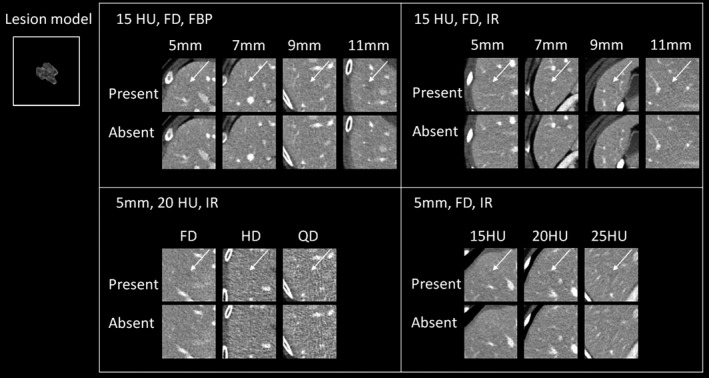
Figure 2.

Example images of lesion‐present and lesion‐absent cases (only the middle image out of the five consecutive images for each trial is shown) across different experimental conditions. The inset image illustrates the middle slice (zoom reconstruction) of the base lesion model used in this study. Upper left: lesion size was varied across 5, 7, 9, and 11 mm, lesion contrast was 15 HU, computed tomography (CT) images were acquired at full routine radiation dose (FD) with the weighted filtered back projection. Upper right: the experimental condition was similar to that in upper left, except that CT images were reconstructed using iterative reconstruction (IR). Bottom left: lesion size was 5 mm, lesion contrast was 20 HU, CT images were acquired with IR, but radiation dose was varied across full, half and quarter of routine dose level (FD, HD, & QD, respectively). Bottom right: lesion size was 5 mm, CT images were acquired with IR at full radiation dose, but lesion contrast was varied across 15, 20, and 25 HU. The arrows indicate the lesion locations.
