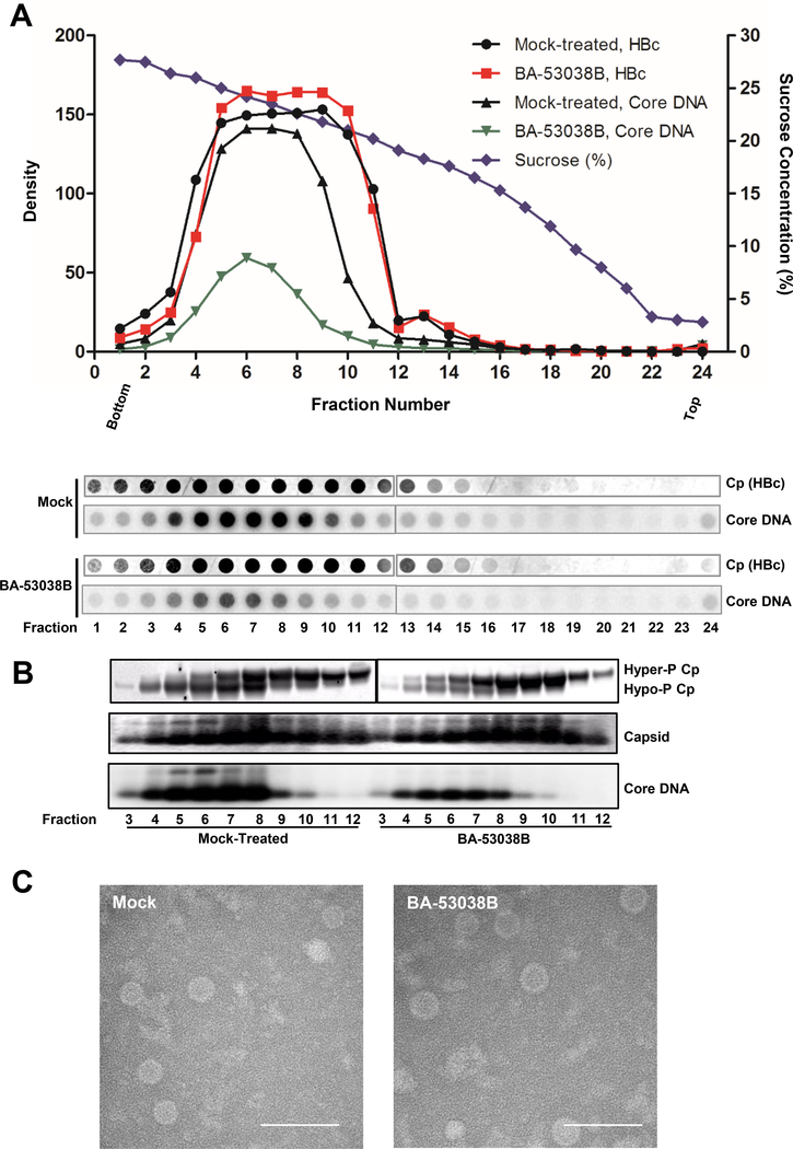
Fig. 6. Physical properties of capsids derived from BA-53038B treated cells.
(A) AML12HBV10 cells were cultured in the absence of tetracycline and treated with 5 μM of BA-53038B. Two days poster treatment, the intracellular capsids were sedimented on a 15% to 30% sucrose. Total of 24fractions were collected from the bottom. 1/30 volume of each fraction was spotted on Nylon membrane for detection of HBV core protein (HBc) with Dako antibody followed by detection of minus strand HBV DNA using HBV riboprobe. (B) HBV capsids from fractions 3–12 were purified by ultracentrifugation and subjected to Western blot to detect core protein with antibody HBc-170A and analyses of capsids and their associated HBV DNA in 1.8 % native agarose gel assay. (C) Electronic microscopic graphs of the capsids purified from AML12HBV10 cells in presence and absence of 5 μM of BA-53038B treatment. Scale bar is 100 nm.

