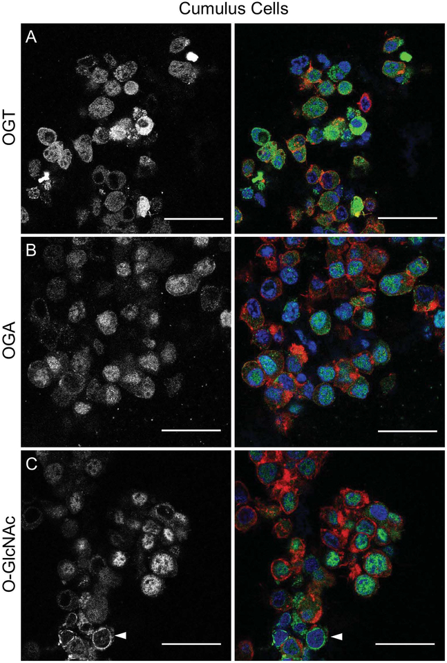
Figure 3. OGA, OGT, and O-GlcNAcylated proteins are expressed in bovine cumulus cells.
Cumulus cells were isolated from bovine COCs collected from antral follicles and analyzed by immunofluorescence and confocal microscopy for (A) OGT, (B) OGA and (C) O-GlcNAc. The gray scale images (left) show the localization of each specific marker, and the merged images (right) show DNA in blue, F-actin in red, and the marker of interest (OGT, OGA, O-GlcNAc) in green. Single optical sections are shown. The arrowhead in (C) highlights O-GlcNAcylated proteins that localize to the nuclear envelope. This experiment was repeated three times with pools of cumulus cells and representative images are shown. The scale bars are 25 μm.

