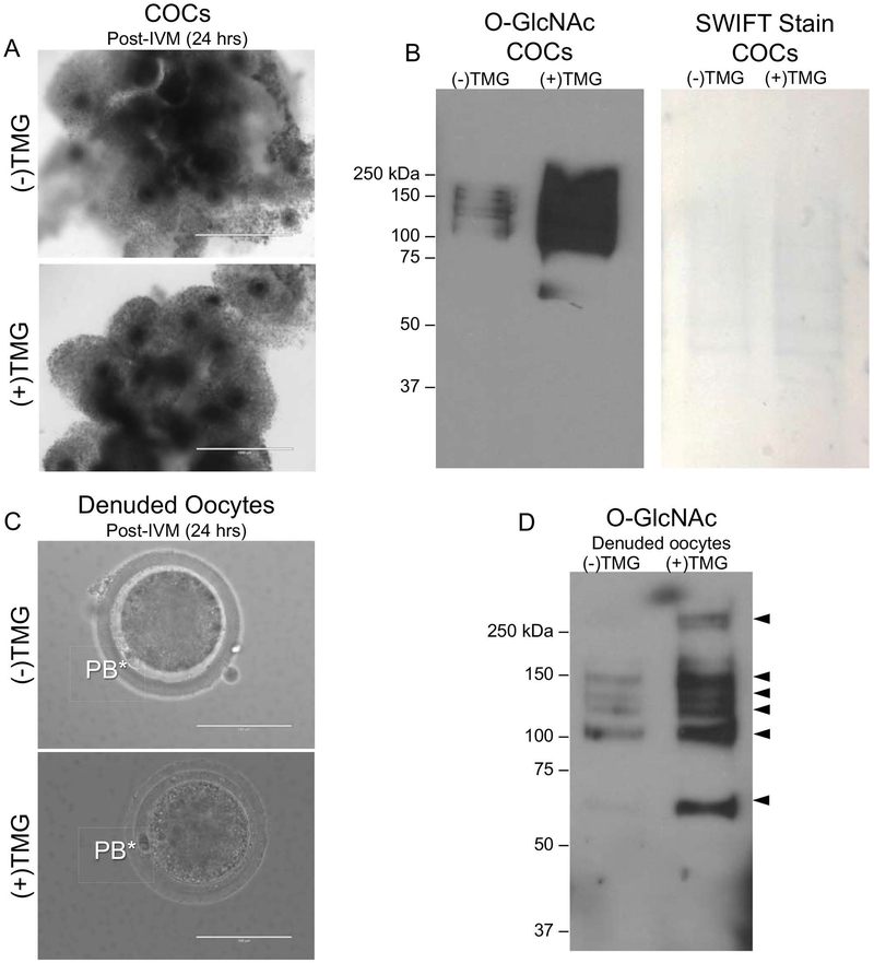
Figure 6. TMG-treatment during in vitro maturation increases O-GlcNAc levels in the bovine COC and oocyte.
(A) Morphology of COCs following +/− treatment with 50 μM TMG for the duration of IVM (24 hours) is shown. Note the equivalent degree of cumulus expansion in both groups. (B) Immunoblot analysis of O-GlcNAc levels in protein extracts from intact bovine COCs treated with 0 μM, and 50 μM for was performed. Protein extracts from 15 COCs were loaded per lane. Swift staining (right) was used to visualize total protein. (C) Representative images of MII eggs isolated from COCs following IVM +/− 50 μM TMG are shown. The asterisks mark the polar bodies (PB). (D) Immunoblot analysis of O-GlcNAc levels in protein extracts from gametes that were stripped from cumulus cells following IVM +/− 50 μM TMG was performed. Protein extracts from equal number of denuded gametes (N = 15) were loaded per lane. All experiments were repeated three times and representative images are shown. The scale bars in (A) are 1 mm and in (C) are 100 μm.

