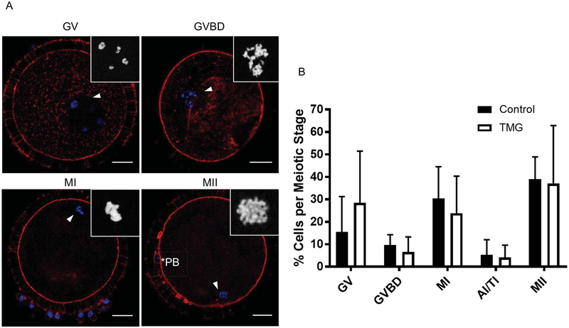
Figure 7. TMG treatment of bovine COCs during in vitro maturation does not impact meiotic progression.
The meiotic stage of oocytes following IVM +/− 50 μM TMG was scored by assessing chromatin configuration via immunostaining and confocal microscopy. (A) Representative images of oocytes at the various meiotic stages are shown (GV; germinal vesicle intact, GVBD; germinal vesicle breakdown, MI; metaphase of meiosis I, and MII; metaphase of meiosis II). DNA is shown in blue and F-actin in red. The arrowhead marks the chromatin that is further magnified in the inset. The asterisks mark the polar body (PB). Single optical sections are shown except for insets where projections are shown. (B) Meiotic progression in each treatment group was plotted and no significant differences were observed. Scale bars in (A) are 25 μm. Meiotic progression data were compiled from nine experimental replicates and included a total of 120 control COCs and 99 TMG-treated COCs.

