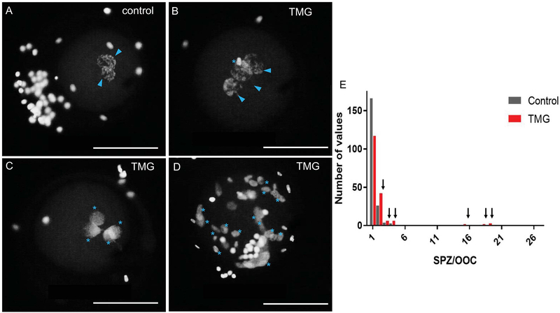
Figure 8. TMG treatment of bovine COCs during in vitro maturation results in zygotic defects.
18 hours post-insemination, zygotes derived from oocytes that were matured +/− 50 μM TMG were fixed to visualize DNA to assess sperm penetration, sperm decondensation, and pronuclear formation. Representative images of (A) control zygotes with two pronuclei and those from (B-D) TMG treatment are shown. The blue arrowheads highlight pronuclei while the blue asterisks highlight multiple sperm in various states of decondensation. The zygote in (D) illustrates an extreme cases of polyspermy. In all images, the nuclei on the outside of the zygote are from lingering cumulus cells. Scale bars are 100 μm. (E) A histogram showing the frequency distribution of the number of spermatozoa per oocyte in zygotes derived from either TMG-treated (red bars) or control oocytes (grey bars). Arrows highlight the increased number of TMG-treated oocytes in all cases of polyspermy. For these experiments, >200 zygotes in each treatment group were analyzed across three replicates.

