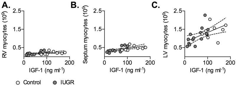
Figure 5. Circulating IGF-1 levels predict cardiomyocyte number in Control and IUGR fetal sheep.
Following significant correlation within IUGR fetuses (P<0.05), regressions on separate treatment groups were found to be not significantly different, and a single regression was fit between circulating IGF-1 levels and A) RV myocyte number (Y=0.0006741X+0.1393, r2=0.1747, P=0.0529), B) septum myocyte number (Y=0.001819X+0.2649, r2=0.4092, P=0.0013), and C) LV myocyte number (Y=0.005212X+0.805, r2=0.3177; P=0.0051). Linear regression±95% confidence intervals are plotted with raw data.

