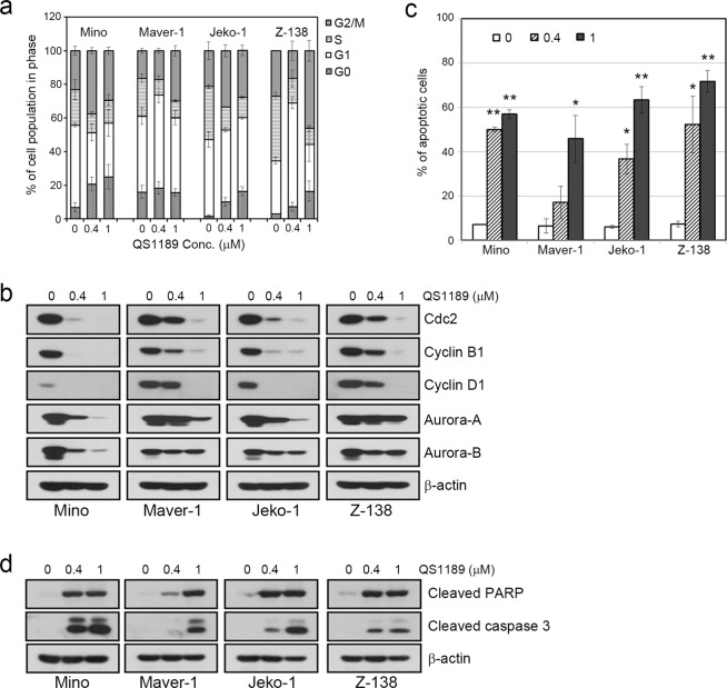
Figure 3.
Induction of cell cycle arrest and apoptosis by QS1189 in MCL cells. (a) MCL cells were treated with QS1189 for 24 h prior to cell cycle analysis by propidium iodide (PI) staining. The experiment was repeated in triplicate. (b) G2/M related protein expression was detected by immunoblotting. (c) MCL cells were treated with QS1189 for 24 h, and then harvested and stained with Annexin V–FITC and PI. The percentage of apoptosis was the sum of the early apoptosis and late apoptosis population. Results are shown as the mean ± standard deviation of three independent experiments. *P < 0.05; **P < 0.005 compared to the control group. (d) Cleaved PARP and caspase-3 levels were detected by immunoblotting.

