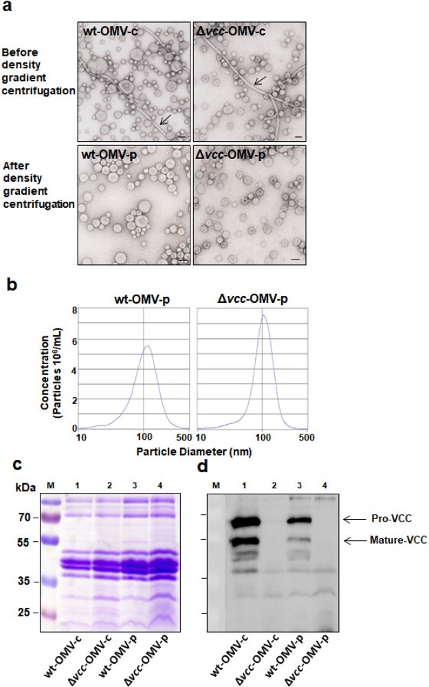
Figure 1.
OMVs subjected to density gradient centrifugation exhibit high purity. (a) Transmission electron microscopy analysis of OMV preparations from wildtype V. cholerae V:5/04 (wt-OMV) and its VCC deletion mutant (Δvcc-OMV) before (wt-OMV-c and Δvcc-OMV-c) and after (wt-OMV-p and Δvcc-OMV-p) density gradient centrifugation. Arrows indicate flagellin filaments. Bars in the lower right of micrographs indicate 100 nm. (b) Nanoparticle tracking analysis of density gradient centrifugation purified OMVs from wildtype V. cholerae V:5/04 (wt-OMV-p) and its VCC deletion mutant (Δvcc-OMV-p). (c) Coomassie-brilliant-blue-stained SDS-PAGE gel and (d) immunoblot analysis of the same gel using polyclonal anti-VCC antiserum. Samples analysed: wt-OMV-c (lane 1), Δvcc-OMV-c (lane 2), wt-OMV-p (lane 3) and Δvcc-OMV-p (lane 4). Molecular weight markers were run in lane M. Molecular weights of the markers are given in kDa to the left of the gel in (c). Arrows in (d) indicate the position of the pro-VCC and the mature VCC protein, respectively. The full-length SDS-PAGE gel and its anti-VCC immunoblot are shown in Supplementary Fig. S1.

