Figure 1.
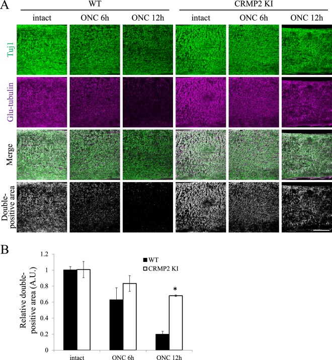
Suppression of microtubule depolymerization in the optic nerves in CRMP2 KI mice. (A) Representative images of the double antibody staining for neuron-specific class III β-tubulin (Tuj1, green) and Glu-tubulin (magenta), which are abundant in the polymerized microtubules. The double-positive areas were remarkably reduced after ONC in wild type mice, indicating microtubule depolymerization. However, the double-positive areas were significantly preserved in those from the CRMP2 KI mice. Scale bar: 100 µm. (B) Relative quantification of the double-positive areas in the optic nerves of wild type and CRMP2 KI mice at 6 and 12 h after ONC. The data were normalized to the double positive areas on the intact side at 12 h after ONC from the wild type. n = 3 mice for each genotypes. *p < 0.05. Statistical analysis was performed using one-way ANOVA, followed by Tukey’s multi-comparison test. Data are represented as. mean ± SEM. h, hours.

