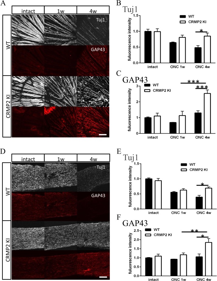
Figure 3.
Immunohistochemical analysis of retina and optic nerve after ONC. (A) Immunohistochemical images of RGC axon layer of the retina with antibodies for Tuj1 and GAP43. (B,C) Quantification of Tuj1 (B) or GAP43 (C) positive area of wild-type (WT) and CRMP2 KI samples after ONC. (n = 4, *p < 0.05 **p < 0.01. Scale bar = 100 µm. (D) Images of immunohistochemistry of optic nerve with antibodies for Tuj1 and GAP43 in longitudinal sections. (E,F) Quantification of Tuj1 (E) or GAP43 (F) positive area of wild-type and CRMP2 KI samples after ONC. (n = 4 mice, *p < 0.05. Scale bar = 100 µm.

