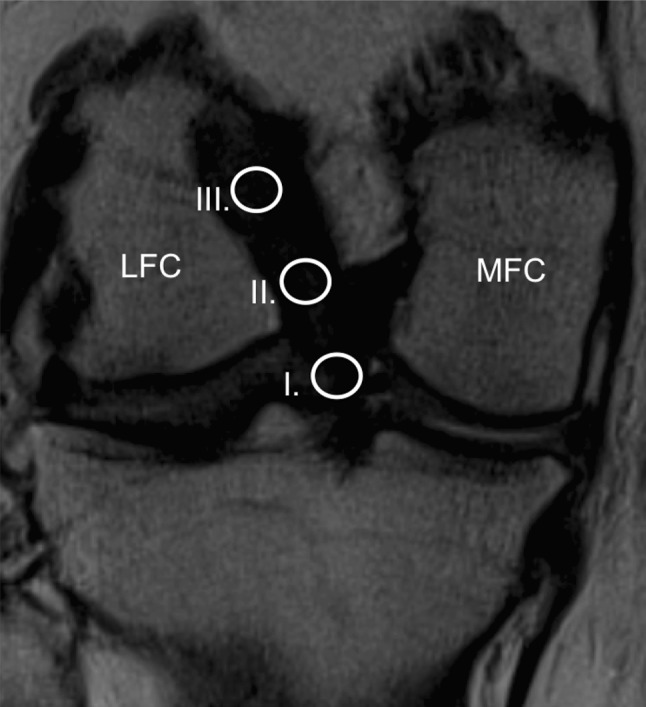
Fig. 1.

Coronal oblique magnetic resonance imaging scan of a right knee showing the position of the three regions of interest (area of the circle = 0.2 cm2), including a (I) distal, (II) middle and (III) proximal site of the ACL. LFC lateral femoral condyle, MFC medial femoral condyle
