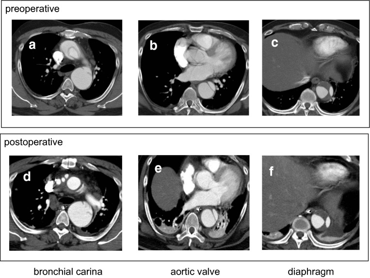
Fig. 4.
CT scan of a patient treated with TAR without FET before 2015. Preoperative image (a–c), and postoperative image before discharge from the hospital (d–f) at the level of bronchial carina (a, d), aortic valve (b, e), and diaphragm (c, f), respectively. Note that, although the elephant trunk was inserted in the descending aorta, the false lumen is patent and the true lumen expansion is not observed at each level of downstream aorta

