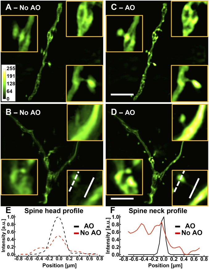
Fig. 1.
AO is essential for SIM imaging in brain tissue. (A–D) Images of dendrites at a depth of 25 m in a cortical slice of a Thy1-GFP line M mouse (A and B) without and (C and D) with AO. (Scale bars: 5 m; Inset widths: A and C, 3 m; B and D, 2 m.) (E and F) Line profiles of (E) a spine head and (F) a spine neck with and without AO as identified by the lines in B and D. Images were normalized to the AO condition.

