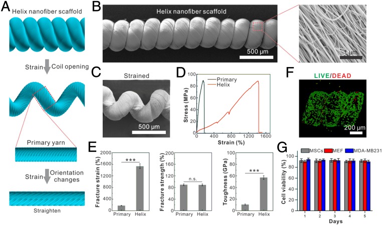
Fig. 1.
Fabrication and characterization of helix microtissue fiber. (A) Schematic illustration of the structure of hierarchical helix nanofiber yarn. (B and C) Scanning electron micrographs of an unstrained (B) and a strained (C) hierarchical helix nanofiber yarn. (D) Stress–strain curve of helix nanofiber yarn shows that the helix scaffold can be stretched up to 15 times its initial length under a stress more than 80 MPa. (E) Quantifications of fracture strain, fracture strength, and fracture toughness show that the helix scaffold’s toughness is 6 times larger than the primary yarn by elevating fracture strain rather than facture strength. ***P < 0.001. n.s., not significant. (F and G) Image (F) and quantification (G) of LIVE/DEAD staining of seeded MEFs show that three different types of cells maintain high cell viability on the helix scaffold during 5 d of culturing.

