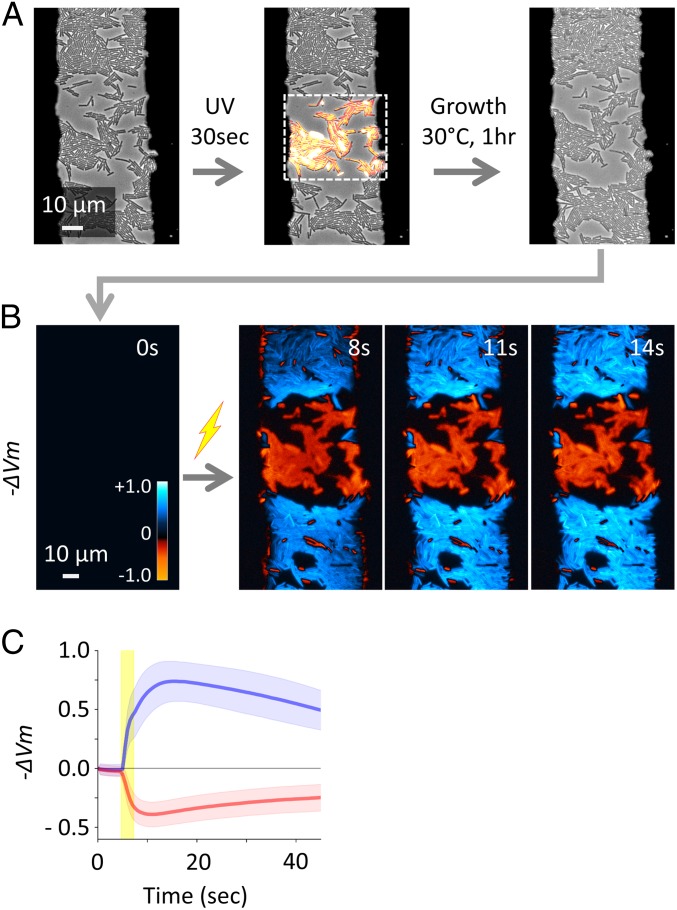
Fig. 2.
UV-V irradiation makes B. subtilis cells respond to an electrical stimulus in the opposite direction. (A) Phase-contrast microscopy image shows WT B. subtilis cells within the electrode gap. A rectangular region indicated by the dashed line within the field of view was irradiated by UV-V light. Growth was suppressed in the UV-V–irradiated region, while cells outside of the UV-irradiation region replicated. (B) The region shown in A was treated with an electrical stimulus. −ΔVm was calculated from ThT fluorescence [log(FThT/FThT,R)] and shown with the colormap in the panel. To an identical electrical stimulus, unperturbed cells hyperpolarized (blue) and UV-V–irradiated cells depolarized (red). (C) Mean (thick lines) and SD (shaded color) of −ΔVm for cells in unperturbed (blue) and UV-V–irradiated (red) regions.

