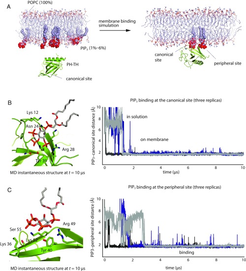
Fig. 1.
Spontaneous binding of the PH–TH module to the membrane. (A) Cartoon illustration of a membrane-binding simulation setup (see SI Appendix, Methods for details). (B, Right) Minimum distance between atoms in PIP3 lipids and in the canonical binding site for three independent membrane-binding simulations. (B, Left) A structure taken from one of these simulations (t = 10 μs) showing a PIP3 molecule bound at the canonical site. (C, Right) Minimum distance between atoms in PIP3 lipids and in the peripheral binding site for three independent membrane-binding simulations. (C, Left) A structure taken from one of these simulations (t = 10 μs) showing a PIP3 molecule bound at the peripheral site.

