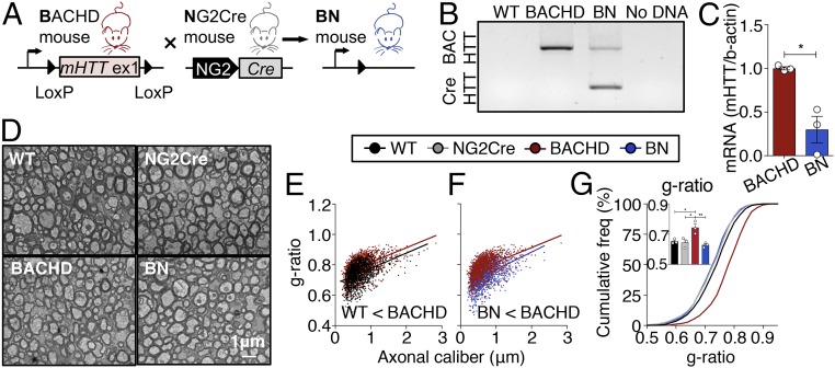
Fig. 1.
OPC-intrinsic effects of mHTT cause myelination abnormalities in HD mice. (A) Schematic representation of Cre-mediated genetic reduction of mHTT expression in OPCs (NG2+ cells) in BACHD mice. (B) PCR analysis confirmed the excision of human mHTT exon 1 in the cortex of BN mice. (C) mHTT mRNA levels are reduced in purified OPCs in BN mice at P6–P7. n = 3/genotype (P = 0.0100, t = 4.601, df = 4). (D) EM images of myelinated axons in the CC at 12 mo of age. (Scale bar, 1 μm.) (E–G) Higher g-ratios (thinner myelin sheaths) in BACHD mice are rescued in BN mice. n = 3/genotype; ∼300 axons were quantified per animal. Data show means ± SEM; *P < 0.05, **P < 0.01; two-tailed Student’s test in C and one-way ANOVA followed by Tukey’s test in G.

