Fig. 4.
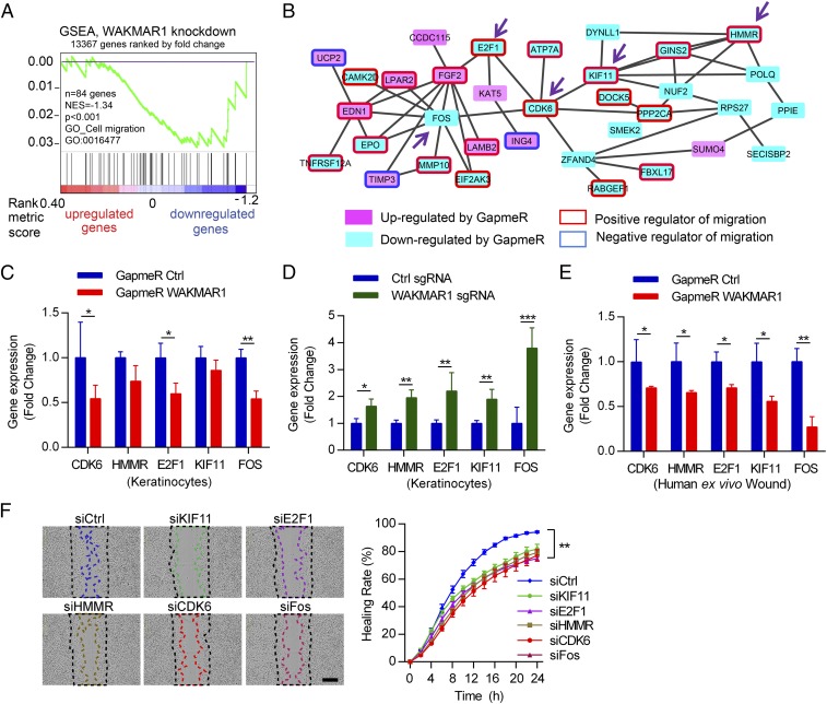
WAKMAR1 regulates a gene network mediating its promigratory function in keratinocytes. Microarray analysis was performed in human keratinocytes with WAKMAR1 knockdown (n = 3). (A) GSEA evaluated enrichment for the cell migration-related genes in the microarray data. NES, normalized enrichment score. (B) Functional protein association network was identified by STRING APP in Cytoscape software among the genes regulated by WAKMAR1 (absolute fold change ≥ 1.3, P < 0.05). Genes up- or down-regulated by WAKMAR1 GapmeR are colored in pink or cyan, respectively. Genes previously reported to promote or inhibit cell migration are highlighted with red or blue frames, respectively. The expression of CDK6, HMMR, E2F1, KIF11, and FOS was analyzed by qPCR in keratinocytes transfected with WAKMAR1 GapmeRs (C), or with CRISPR/Cas9-SAM plasmids (D), and in human ex vivo wounds treated with WAKMAR1 GapmeRs (E). (F) Scratch wound assay of keratinocytes transfected with siRNAs specific to KIF11, E2F1, HMMR, CDK6, or FOS (n = 8). (Scale bar, 300 μm.) *P < 0.05; **P < 0.01; ***P < 0.001 by unpaired two-tailed Student’s t test (C–E) or two way-ANOVA (F). Data are presented as mean ± SD and are representative of at least two independent experiments.

