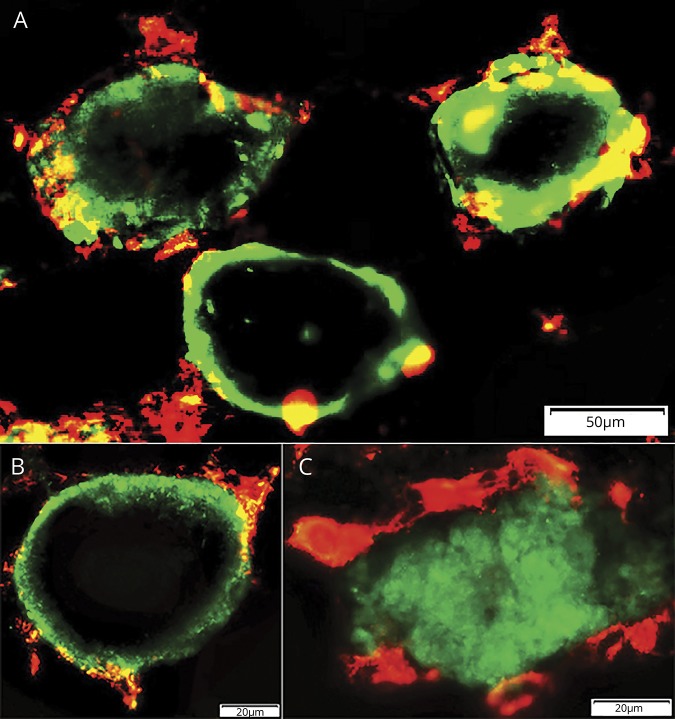
Figure 2. Histiocytic cells: relationship to C5b-9 stained muscle fiber cytoplasm.
(A, B). Histiocytes stained for HAM56 (red), a pan-histiocyte marker, are present around, and within, damaged, C5b-9 stained (green) cytoplasm in the periphery of muscle fibers. Bars = 50 μm (A) and 20 μm (B). (C) Large histiocytes stained for HAM56 (red) surround a necrotic muscle fiber with diffusely C5b-9 stained (green) cytoplasm. This diffuse pattern of C5b-9 staining of the cytoplasm of necrotic muscle fibers is the typical pattern observed in most necrotic fibers in other disorders. Bar = 20 μm.

