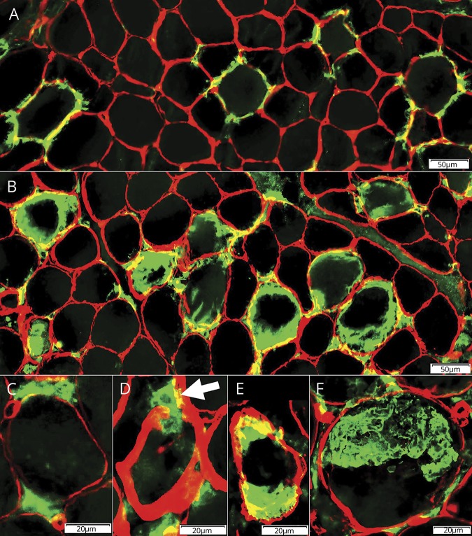
Figure 3. Histiocytic cells: relation to collagen 4–stained muscle fiber basal lamina.
(A) Early pathology: histiocytes stained for S100 (green) are present outside, and over, collagen 4–stained muscle fiber basal lamina (red). Bar = 50 μm. (B) Later pathology: histiocytes stained for S100 (green) are present inside the collagen 4–stained muscle fiber basal lamina (red), and within areas occupied by muscle fibers, but not within the central regions of these areas. Bar = 50 μm. (C) Histiocytes stained for CD68 (green) are present around collagen 4–stained muscle fiber basal lamina (red). Bar = 20 μm. (D) Histiocyte stained for CD163 (green) (arrow) extends through the collagen 4–stained muscle fiber basal lamina (red). Bar = 20 μm. (E) Large histiocytes stained for CD163 (green) inside the collagen 4–stained muscle fiber basal lamina (red). Bar = 20 μm. (F) Large region of 1 or several histiocytes stained for CD163 (green) replacing area of a muscle fiber inside the collagen 4–stained muscle fiber basal lamina (red). Bar = 20 μm.

