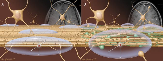
Figure 2. Theoretical basis of diffusion imaging.
(A) Water diffusion along intact axonal fiber tracts in intact brain tissue. (B) Dispersed water diffusion in brain tissue with demyelination and axonal injury. The arrows indicate the primary, second, and third eigenvectors of the diffusion tensor. Reprinted with permission, Cleveland Clinic Center for Medical Art & Photography © 2004–2018. All Rights Reserved.

