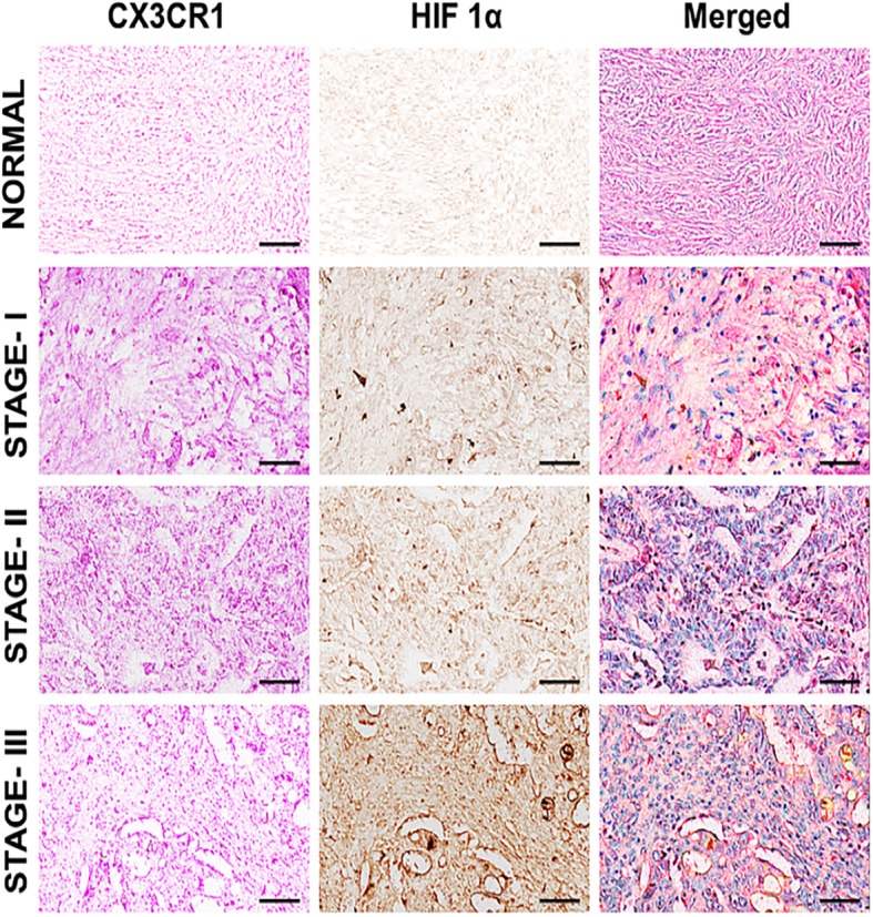
Fig. 1.

CX3CR1 and HIF-1α expression by OvCa tissues: Ovarian tissues from normal and various cancer stages [well-differentiated (Stage I), moderately differentiated (Stage II), and poorly differentiated (Stage III)] were stained with anti-CX3CR1 and anti-HIF-1α antibodies. Magenta (AP) color shows CX3CR1, and Brown (DAB) color shows HIF-1α staining. An Aperio ScanScope CS system with a 40X objective captured digital images of each tissue. Representative cases are immuno-intensities of CX3CR1 and HIF-1α using image analysis Aperio ImageScope v.6.25 software
