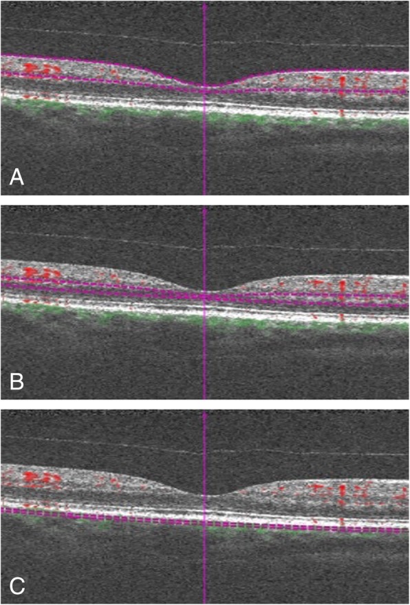
Fig. 1.

B-scan pictures showing the automatic segmentation of the vascular plexuses. The superficial capillary plexus (a) is automatically segmented using as inner boundary the ZILM (internal limiting membrane layer) and as outer boundary the ZIPL (inner plexiform player). The deep capillary plexus (b) inner boundary is ZIPL, while the outer is ZOPL (outer plexiform layer). The OPL is estimated as ZRPE (retinal pigment epithelium layer) - 110 μm, and the ZIPL is calculated as: ZILM + 70%*(TILM-OPL). The choriocapillaris (c) inner boundary is ZRPE + 29 μm, and the outer is ZRPE + 49 μm. Violet dashed lines designate the boundaries, while red and green spots respectively represent the perfusion above and below the RPE
