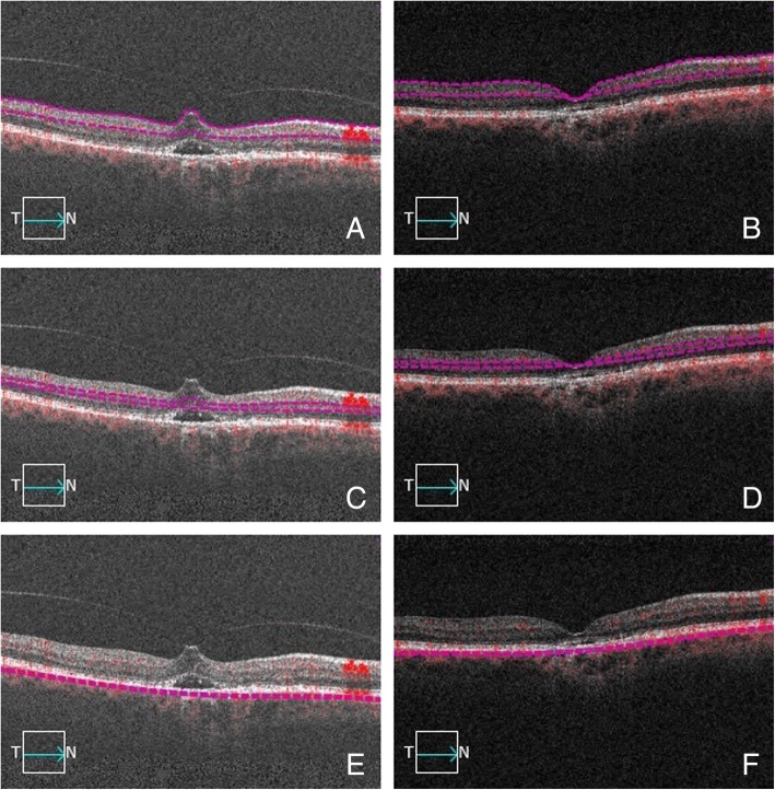
Fig. 3.
B-scan pictures showing the automatic segmentation of the three vascular plexuses before and after intravitreal ocriplasmin injection in a case achieving complete anatomical resolution. Violet dashed lines designate the correctly-segmented boundaries of the superficial capillary plexus (a), deep capillary plexus (c) and choriocapillaris (e) before ocriplasmin injection. Focal vitreomacular traction with neuroepithelial detachment is evident. Compete vitreomacular traction release is evident after injection (respectively b, d, f)

