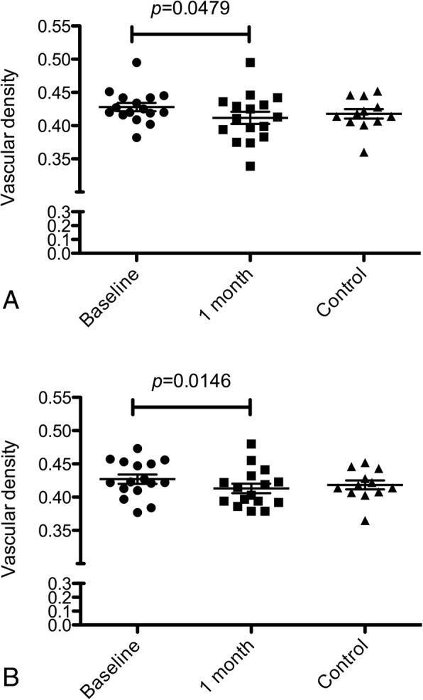
Fig. 5.

Box-and-whiskers plots of capillary perfusion density in the superficial capillary plexus (SCP). The plots show the perfusion density in the retinal SCP of eyes with idiopathic vitreomacular traction at baseline and at 1 month after ocriplasmin injection, compared with healthy controls. Perfusion density was calculated respectively in the 3 × 3 mm (a) and in the 6 × 6 mm (b) scans
