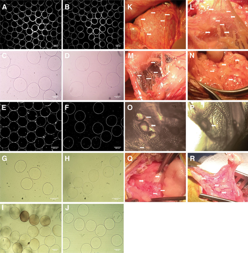
FIGURE 1.

Images of the original empty microbeads preimplantation and appearance of retrieved microbeads postexplant, together with the gross surgical appearance at the specific time points. Set of the preimplantation microbeads used for the NHP implants are shown (A) without CXCL12 (−) and (B) with CXCL12 (+). Size and shape of the microbeads were very similar between the CXCL12 (−) and CXCL12 (+) samples, magnification ×4. Images of the microbeads postexplant at day 30 are shown (C) without CXCL12 (−) and (D) with CXCL12 (+). Similar images shown in A–B and E–F were used by University of California Irvine and their software program for the microbeads size and shape evaluation. Explanted microbeads at day 90 are shown (G) without CXCL12 (−) and (H) with CXCL12 (+). Microbeads measured 500–530 μm in diameter, magnification ×10. Explanted microbeads at day 180 are shown (I) without CXCL12 (−) and (J) with CXCL12 (+). Microbeads explanted from the CXCL12 (−) NHP were in the range of 480 ± 43 μm, and some microbeads appear to have a pigmentation with changed surface but without obvious cell infiltration. Microbeads from the CXCL12 (+) NHP were in the range of 485 ± 12 μm, magnification ×10. Images from the surgical appearance of microbeads (shown with arrows) at days 30, 90, and 180 demonstrate visible numerous concentrations of microbeads within the peritoneal cavity of NHPs (K) without CXCL12 (−) and (L) with CXCL12 (+) at day 30. M, Image of the omental tissue with CXCL12 (−) NHP demonstrates the cluster of microbeads (N) with CXCL12 (+) NHP at day 90. Microscopic images of the tissues with embedded empty microbeads from the CXCL12 (−) explant at day 180 are shown with arrows in O–P, demonstrating the “vesicle-like” pouch around the single microbeads with a visible vascularization, and tissues with embedded empty microbeads from the CXCL12 (+) are seen in Q and R. NHP, nonhuman primate.
