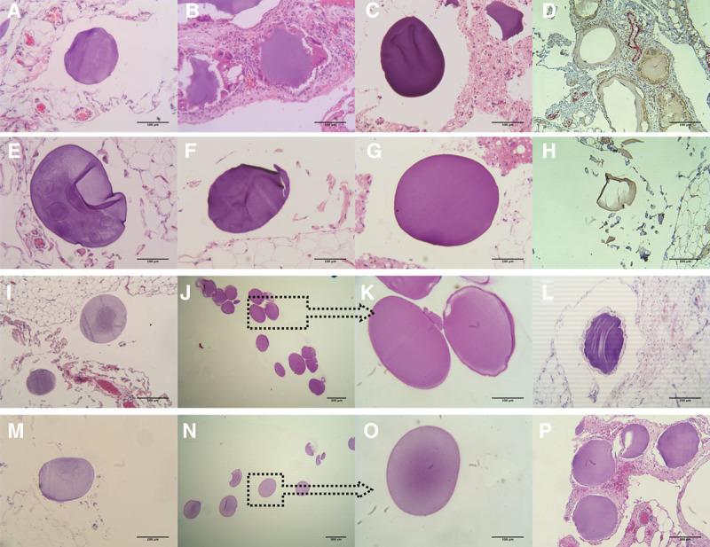
FIGURE 2.

Histopathology of the explanted empty microbeads without CXCL12 (−) and with CXCL12 (+) from days 30 to 180. Samples of the microbeads at day 30 are shown in A–H. A, Tissues with embedded microbeads explanted from the NHP without CXCL12 (−). B, Fibrosis of the tissues with microbeads from the CXCL12 (−) NHP. C, Microbead sample found in the aspirate debris. D, IHC staining of the tissues from CXCL12 (−) NHP, stained with antifibroblast activation protein alpha (brown color) and with antimacrophage (red color), magnification ×10. E–G, Tissues with embedded microbeads explanted from the NHP with CXCL12 (+). H, IHC staining of the tissues from CXCL12 (+) NHP, with same staining as for the CXCL12 (−) NHP, magnification ×10. Images showing the explanted tissues (I) and free microbeads (J and K) from CXCL12 (−) NHP and explanted tissues (M) and free microbeads (N and O) from CXCL12 (+) NHP, respectively, at day 90. As it can be seen in the panels with free microbeads, there were no visible cell attachments or fibrosis present in these samples, magnification ×4 and ×20. H&E staining of the explanted tissues from CXCL12 (−) NHP with embedded microbeads at day 180 is shown in image (L) and from CXCL12 (+) NHP shown in image (P), magnification ×10. H&E, hematoxylin and eosin; IHC, immunohistochemistry; NHP, nonhuman primate.
