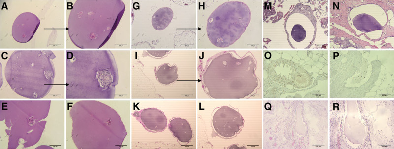
FIGURE 4.

Histopathology on explanted free and embedded microbeads from autologous implants at days 30 and 180. A–F, H&E staining of the recovered free microbeads from the first autograft at day 30. Visible nucleated islets and also some denucleated clusters. E, Visible damage from the recovery or staining process. G and H, H&E staining of the tissue-embedded microbeads from the first autograft at day 30. Image showed no cellularization around the capsule but some denucleation of the islets. I–L, H&E staining of the recovered embedded microbeads from the first autograft at day 180. Visible presence of more denucleated islet clusters, but still without significant cellularization. M and N, Histology of the embedded microbeads from the second autograft at day 30. Images showing no cellularization around the capsules. O and P, IHC staining of the embedded microbeads from the second autograft at day 30. Tissues around microbeads stained with antifibroblast demonstrate the presence of the cells and damage to the microbead structure. Q and R, H&E staining of the recovered embedded microbeads from the second autograft at day 180. There were no detectable islet clusters in these microbead samples. H&E, hematoxylin and eosin; IHC, immunohistochemistry.
