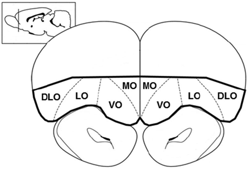
Fig. 2.

Graphical representation of region dissected out for Western Blotting, bolded in black outline. Coronal sections containing the orbitofrontal cortex (OFC) were cut in a cryostat at −20°C using coordinates from Paxinos and Watson (1998) (OFC: AP +4.2 mm to +2.7 mm). Using a scalpel and visual cues from natural boundaries, the OFC was dissected at −20°C. DLO, dorsolateral orbitofrontal cortex; LO, lateral orbitofrontal cortex; MO, medial orbitofrontal cortex, VO, ventral orbitofrontal cortex.
