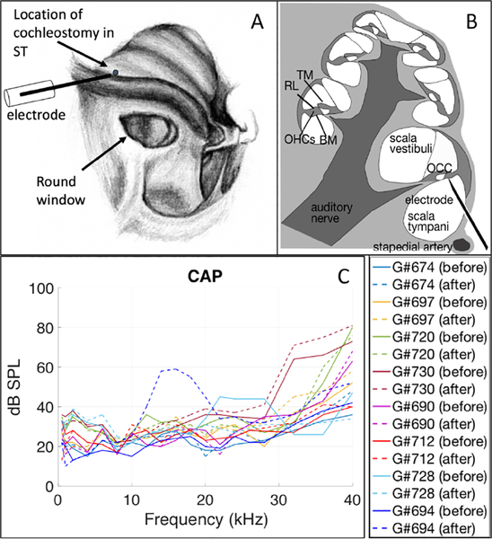
Figure 1:
A) view of the gerbil cochlea from the bulla opening (drawing by Vanessa Cervantes). VOHC responses were measured via a hand-drilled hole (~ 100 μm) in ST in the base of cochlea. Displacement responses were measured through the intact round window membrane. B) Cross-section of the cochlea, showing the electrode positioned close under the organ of Corti complex (OCC) in the first turn. The OCC spirals around the cochlea, and some of its main parts are labeled in the section on the left. TM: tectorial membrane, RL: reticular lamina, BM: basilar membrane, OHCs: outer hair cells. C) Compound action potential (CAP) thresholds before (solid lines) and after (dashed lines) the cochleostomy for eight extracellular voltage experiments.

