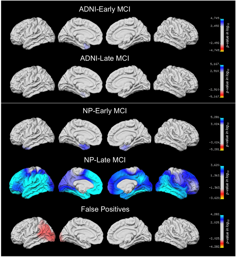
Fig. 5.
T-value surface maps showing regional cortical thickness on the left and right lateral and medial pial surfaces for each group relative to the CN group with FDR correction for multiple comparisons. The cyan/blue shades represent areas where the MCI subgroup has thinner cortex than the CN group, while the red regions represent areas were the subgroup has thicker cortex than the CN group.

