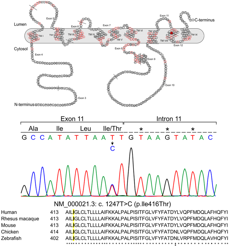
Figure 2.

A: schematic representation of Preselenin 1 in the cell membrane. Residues that have variants known to cause Alzheimer’s disease are represented with a red border [3]. Residue 416 in TM VIII is colored in red. TM: Transmembrane
B: Color chromatogram depicting the missense variant in the PSEN1 gene.
C: Alignment of PSEN1 ortologs (uniprot.org). Residue 416 is highlighted in yellow.
* Residues that are conserved among the 5 species.
• Residues that are conserved at least in 4 species.
: Residues that are conserved at least in 3 species.
