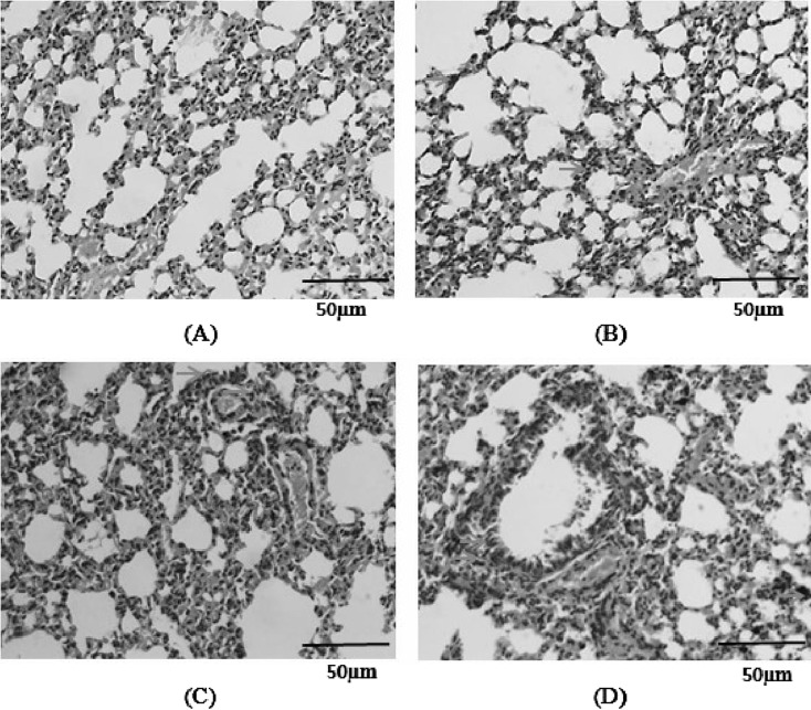
Fig. 5.
Histopathological analysis of the lung in mice exposed to PM, S. aureus, or their combination. Hematoxylin and Eosin (H&E) staining is shown (×400). (A) Control, (B) G1, (C) G2, (D) G3. Yellow arrows indicate leukocytes, red arrows indicate goblet cells, and blue arrows indicate particulates.

