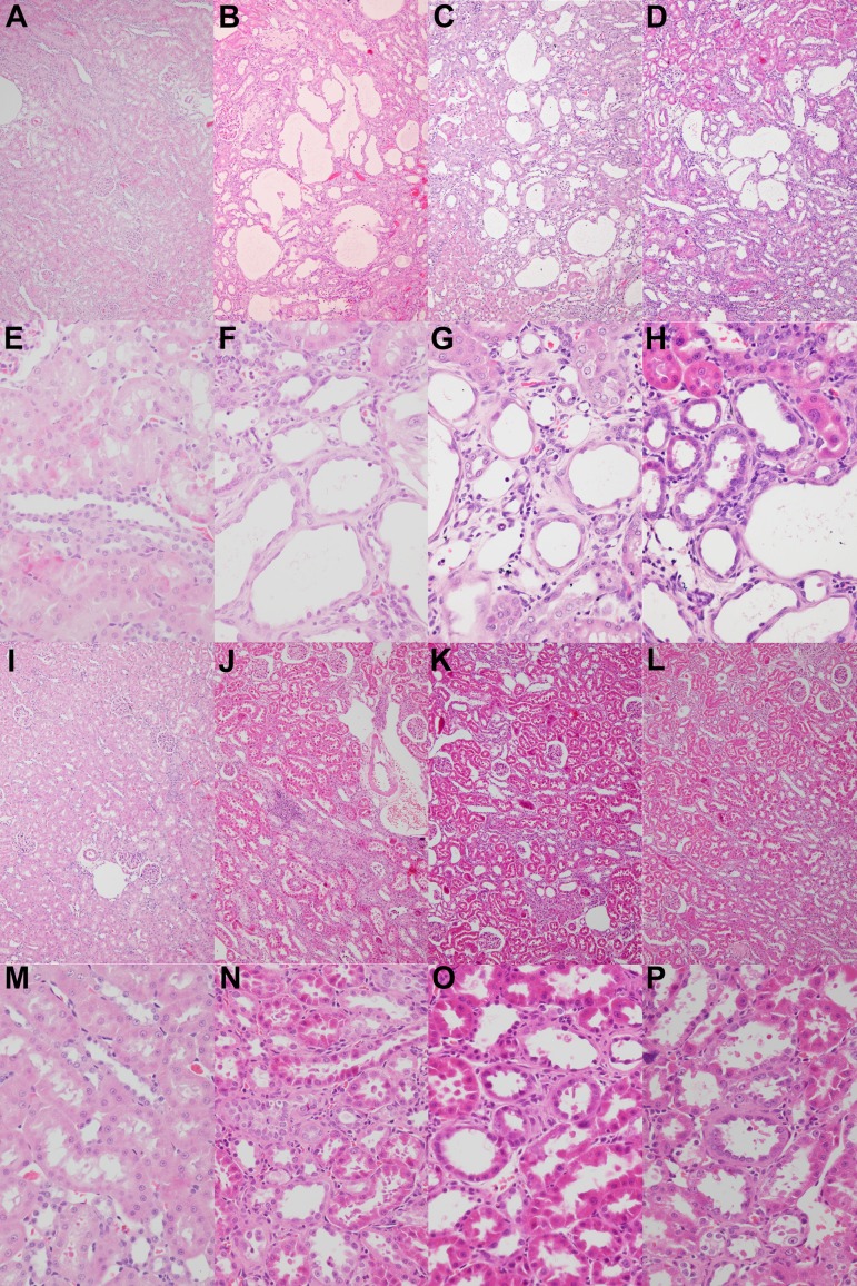
Fig. 2.
Histopathological changes. There was no change in the control group (A and E). In Experiment 1, tubular lesions such as necrosis, degeneration, and luminal dilation; mononuclear cell infiltration; and peritubular fibrosis were prominent in the CIS group (B and F). These histological changes were ameliorated by 300 ppm (C and G) and 1,500 ppm (D and H) DMF and recovered to the control level by 7,500 ppm DMF (I and M). Additionally, in Experiment 2, these changes were dose-dependently ameliorated in the 2,000 ppm (J and N), 4,000 ppm (K and O), and 6,000 ppm (L and P) DMF groups. Hematoxylin and eosin staining. Magnification: ×100 (A–D and I–L) or ×400 (E–H and M–P).

