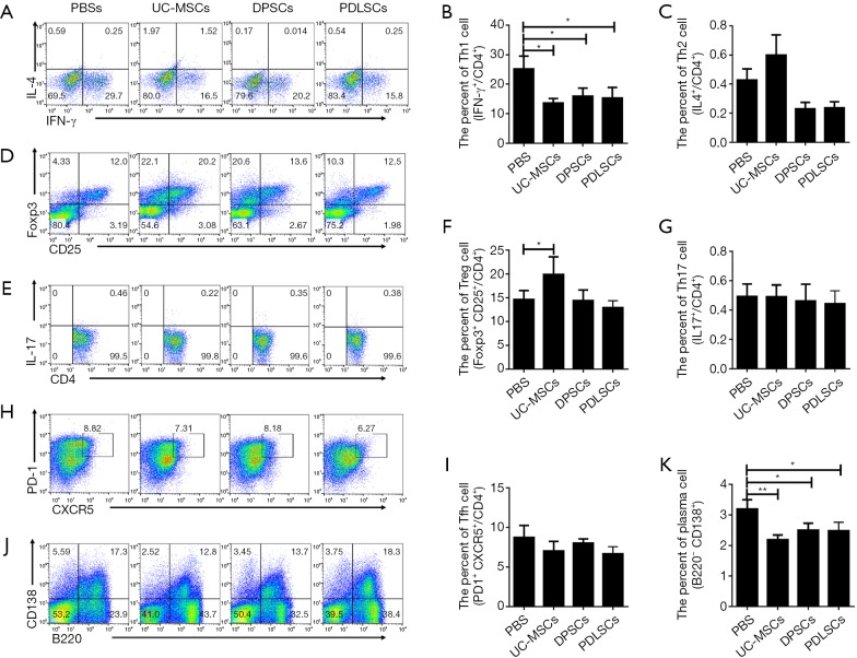
Figure 5.
Flow cytometric analysis of Th1, Th2, Treg, Th17, Tfh and plasma cells in spleens of B6/lpr mice. (A,D,E,H,J) Representative FACS images of CD4+IFN-γ+ (Th1 cells), CD4+IL4+ (Th2 cells), CD4+CD25+Foxp3+ (Treg cells), CD4+PD-1+CXCR5+ (Tfh cells), CD4+IL17+ (Th17 cells) and B220−CD138+ (plasma cells). (B,C,F,G,I,K) Percentages of Th1, Th2, Treg, Tfh, Th17 and plasma cells in the spleen of B6/lpr mice with or without transplantation of MSCs. Data are mean ± SEM. PBS (n=8), UC-MSCs (n=8), PDLSCs (n=8), DPSCs (n=9). *P<0.05, **P<0.01, Kruskal-Wallis test multiple comparison posttest. PBS, phosphate buffered saline; UC-MSCs, umbilical cord mesenchymal stem cells; DPSCs, dental pulp stem cells; PDLSCs, periodontal ligament stem cells.

