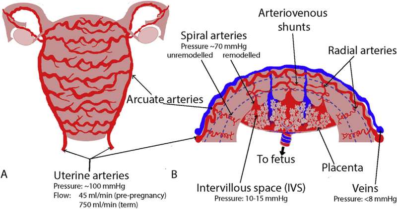
Figure 1:

A schematic of the structure of the uterine vasculature in pregnancy. Blood is primarily delivered to the uterus via the uterine arteries, which branch to the arcuate arteries. The arcuate arteries traverse the uterus in “horizontal rings” (Panel A). The arcuate arteries then branch through the walls of the uterus to radial, basal and spiral arteries, which in pregnancy deliver maternal blood to the site of the placenta where it bathes the placental structures in the intervillous space (Panel B). There are also arterio-venous (A-V) anastomoses (shunt pathways) that connect myometrial radial arteries to the venous side of the uterine vasculature. These anastomoses potentially carry the majority of uterine blood-flow post-delivery. The blue dotted lines in B approximately delineate the myometrium from the endometrium in the placental bed, and the myometrium from the perimetrium near the surface of the uterus.
