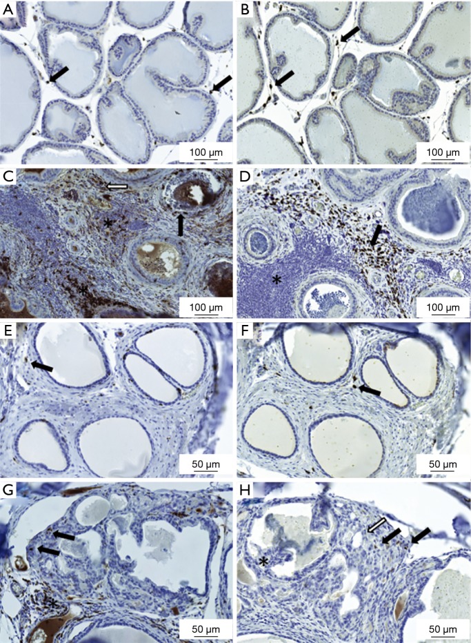
Figure 2.
Presence of macrophages in the prostate and periurethral area. (A-D) Representative figures demonstrating presence of CD68- and CD163-positive macrophages in the lateral prostate lobe of the 18-week treated Noble strain. Only few (A) CD68- or (B) CD163-positive macrophages are present in the placebo rat (arrows); (C) presence of CD68-positive cells in the prostate acini (black arrow), stroma (white arrow) and inside the inflammation infiltrate areas (stars) in the lateral prostate lobe from a hormone-treated rat; (D) representative figure from a hormone-treated rat with extent CD163-positive cells in the prostatic stroma (arrow) but not in the inflammation infiltrate areas (star) of the lateral prostate lobe; (E-H) CD68- and CD163-positive macrophage staining in the periurethral area; (E) only few CD68-positive cells or (F) CD163-positive are seen in the healthy prostate tissue around the prostatic ducts (arrows) in the placebo-treated rat; (G) cancerous areas in the periurethral space around the prostatic ducts with infiltration of CD68-positive macrophages in the connective tissue (star) around the cancerous area and few cells inside the cancerous region in the hormone-treated rat (arrows); (H) hormone-treated rat with PIN-like lesions (star) and cancerous area (white arrow) in the periurethral space around the prostatic ducts and only few CD163-positive cells visible around that area (black arrows). (A-D) 150× magnification, (E-H) 200× magnification.

