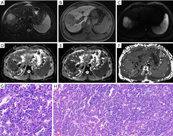
Figure 4.
MR images of a 31-year-old male patient with a pathologically verified HCC of Edmondson-Steiner grade III and MVI positive. The patient suffered intrahepatic recurrence at 8 months after tumor resection. A 6.3 cm tumor in right lobe of the liver shows hyperintensity on T2-weighted image (A), hypointensity on 20-min hepatobiliary phase (B) and restricted diffusion on the diffusion-weighted image with a b-value of 700 s/mm2 (C). ADC (D) and MD maps (E) shows lower signal intensity compared with that of liver parenchyma. MK map (F) shows higher signal intensity of tumor compared with that of background liver parenchyma. The calculated mean values of ADC, MD and MK for the HCC were 1.18×10⁻³ mm2/s, 1.24×10⁻³ mm2/s, and 0.96, respectively. The tumor was histopathologically proven to be Edmondson-Steiner III grade with hematoxylin-eosin (HE) staining at 200× magnification (G). The HE staining at 100× magnification (H) showed that increased cellularity, marked variation of nuclear pleomorphism and disorganized distribution of tumor cells with different differentiation degrees. HCC, hepatocellular carcinoma; MVI, microvascular invasion; ADC, apparent diffusion coefficient; MD, mean corrected apparent diffusion coefficient; MK, mean diffusion kurtosis coefficient.

