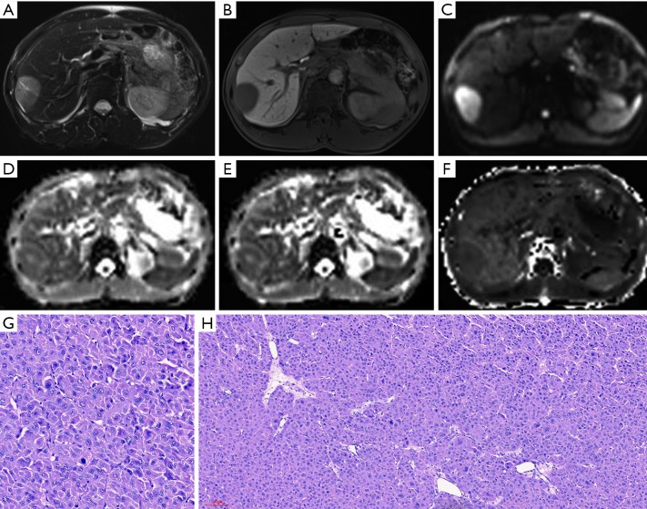
Figure 5.
MR images of a 48-year-old male patient with a pathologically verified HCC of Edmondson-Steiner grade II and MVI negative. The patient did not have tumor recurrence within 1 year. A 4.6 cm tumor in the right anterior hepatic section shows hyperintensity on T2-weighted imaging (A), hypointensity relative to the surrounding liver parenchyma in hepatobiliary phase (B), and restrict diffusion on the diffusion-weighted image with a b-value of 700 s/mm2 (C). ADC (D) and MD maps (E) show slightly higher signal intensity compared with that of liver parenchyma. MK map (F) shows lower slightly lower signal intensity of tumor compared with that of background liver parenchyma. The calculated mean values of ADC, MD, and MK for the HCC were 1.34×10⁻³ mm2/s, 1.60×10⁻³ mm2/s and 0.80, respectively. The hematoxylin-eosin (HE) staining of the tumor at 200 × magnification proved it to be Edmonson-Steiner grade II (G). The HE staining at 100× magnification (H) showed that increased cellularity relatively lowered structural complexity. HCC, hepatocellular carcinoma; MVI, microvascular invasion; ADC, apparent diffusion coefficient; MD, mean corrected apparent diffusion coefficient; MK, mean diffusion kurtosis coefficient.

