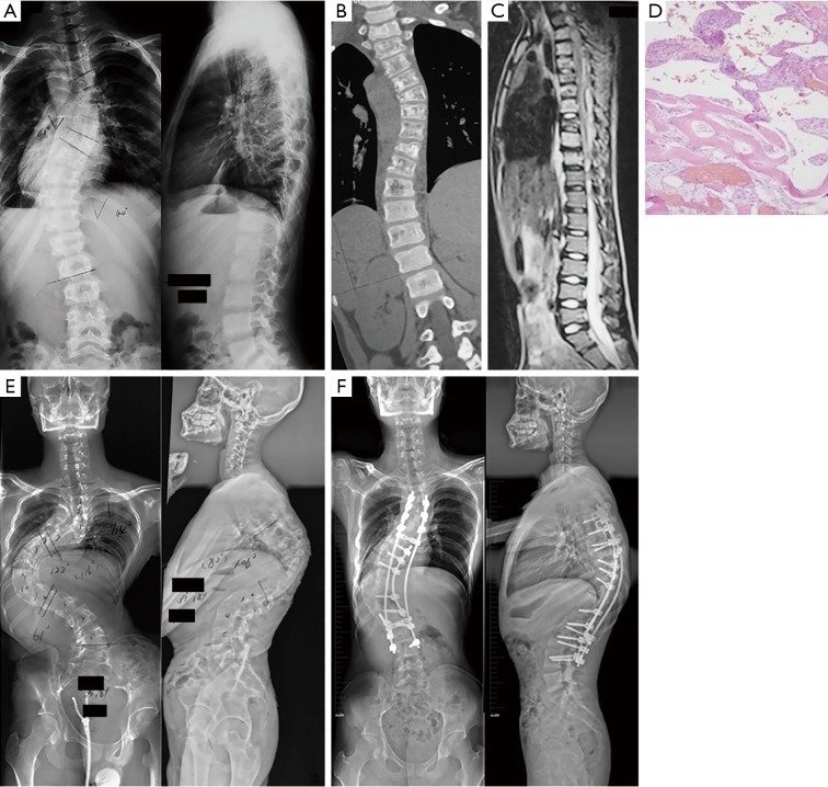
Figure 3.
Patient 4, male, 12 years. (A) Initial X-rays showing C-shaped thoracolumbar scoliosis with a Cobb angle of 61°. (B,C) CT scanning and MRI showing aggressive osteolysis and destructive lesions of the thoracic vertebra. (D) Hematoxylin and eosin staining showing thin-walled blood vessels with hemorrhage and scanty bone (magnification at 10×). (E) Corrective surgery was postponed because of the patient’s extremely low bone mineral density, and was then replaced with Boston bracing. The compensatory thoracolumbar curve increased to 122°, with a severe thoracic kyphosis of 106° and the apex at T10 7 years later. (F) Surgery with satellite rod and screws from T3 to L4 was performed after 2 months of Halo traction, and a significant improvement of the spinal deformity with rigid fusion was achieved. CT, computed tomography; MRI, magnetic resonance imaging.

