Figure 5.

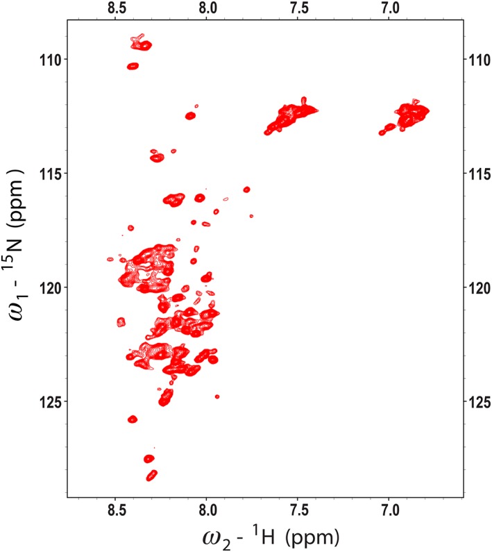
2D 1H,15N HSQC spectrum of [U‐15N]‐RALF1 collected at 600 MHz (1H). The lack of dispersion in the 1H dimension and broad peaks indicate that the peptide is unstructured and aggregated.

2D 1H,15N HSQC spectrum of [U‐15N]‐RALF1 collected at 600 MHz (1H). The lack of dispersion in the 1H dimension and broad peaks indicate that the peptide is unstructured and aggregated.