Figure 6.

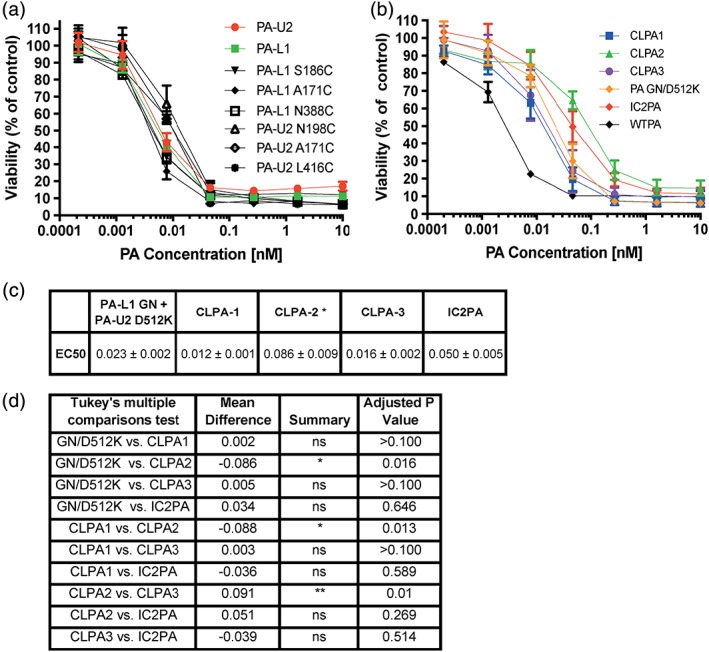
CLPA toxicity on LLC cells. (a, b) LLC cells exhibiting 30% confluence were incubated with serial dilutions of PA variants (0–10 nM) and FP59 (1.9 nM). In (a), cytotoxicities of the monomeric, unconjugated PA‐L1 and PA‐U2 single cysteine variants were assessed. In (b), cytotoxicity of the CLPA variants was compared with WTPA and the two intercomplementing models, IC2PA and PA‐L1 GN + PA‐U2 D512K. In all cases cell viability was measured using an MTT assay after 48 h. (c) Table outlining EC50 (nM) values for PA variant titrations on LLC cells. Errors are displayed as ± error of the mean of at least nine biological replicates. *represents a significant difference in toxicity for the CLPA2 variant as compared to PA‐L1 GN + PA‐U2 D512K, CLPA1 and CLPA3, but no significant difference found as compared to IC2PA. (d) Using GraphPad a one‐way Anova Tukey's multiple comparisons test was completed to compare cytotoxicity of the PA variants to each other in LLC assays. Six biological replicates of calculated EC50 values from experiments containing at least three replicates were used. The **represents a P value of ≤0.0012 and *represents a P value of ≤0.03.
