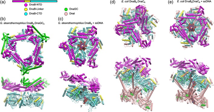
Figure 3.

Cartoon representations of DnaB helicase. (a) Arrangement and color coding of DnaB domains and associated proteins. (b) Orthogonal views of G. stearothermophilus DnaB6.DnaGC3 complex (PDB ID 2R6A). (c) Orthogonal views of G. stearothermophilus DnaB6 in complex with ssDNA (VDW spheres) (PDB ID 4ESV). (d) Orthogonal views of the DnaB6.DnaC6 complex (E. coli) (PDB ID 6QEL). (e) Orthogonal views of the DnaB6.DnaC6 complex with ssDNA (E. coli) (PDB ID 6QEM). Bound nucleoside phosphates are represented as green VDW spheres.
