
Figure 1.
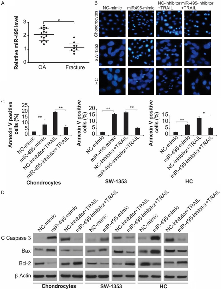
miR-495 promotes apoptosis of primary cultured rat chondrocytes, HC cells and SW1353 cells. A. Relative miR-495 level in clinical specimens (OA = 16, Fracture = 9) was detected by Real-time RT-PCR. *, P < 0.05. B. Indicated cells were treated with NC-mimic, miR-495-mimic, TRAIL + NC-inhibitor or TRAIL + miR-495-inhibitor as indicated. Photomicrographs of the nucleus the cells stained with DAPI. C. Indicated cells were treated with NC-mimic, miR-495-mimic, TRAIL + NC-inhibitor or TRAIL + miR-495-inhibitor as indicated. Cell apoptosis was analyzed by flow cytometry. Data represent the mean ± SD of three independent experiments. **, P < 0.01; *, P < 0.05 (one-way ANOVA with Tukey’s post hoc test). D. Indicated cells were treated with NC-mimic, miR-495-mimic, TRAIL + NC-inhibitor or TRAIL + miR-495-inhibitor as indicated. The protein level of cleaved caspase-3, Bax and Bcl-2 were measured by western blot assay.