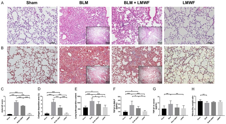
Figure 1.
LMWF alleviated lung fibrogenesis and TGF-β1 expression in a BLM-induced PF mouse model. (A) Representative micrographs of hematoxylin and eosin (H&E) stain for each group on day 28. (B) Masson’s trichrome staining for each group. The blue regions indicated the collagen deposition, which widely distributed in lung interstitial space. Outer original images are × 200 magnification, scale bar = 100 μm; inner original images are × 40 magnification, scale bar = 50 μm. (C and D) Ashcroft score and the collagen deposition area (%) for each group. Data were analyzed using one-way analysis of variance (ANOVA) and 5-7 animals were quantified in each group. (E) Lung hydroxyproline content. (F) Level of expression of TGF-β1 in bronchoalveolar lavage fluid (BALF). (G) Level of expression of TGF-β1 in the serum. (H) Lung wet-to-dry weight ratio (W/D) in each group. The data of (E-H) were analyzed by using ANOVA and are here presented as the mean value ± SD for 8-12 individuals in each group. Significance level was labeled as *P < 0.05, **P < 0.01, ***P < 0.001.

