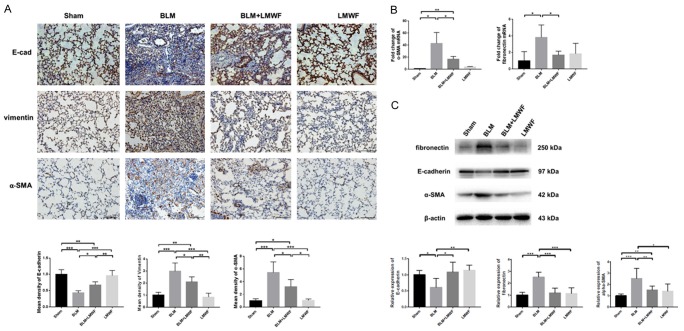
Figure 2.
LMWF attenuated lung EMT phenotype in a BLM-induced PF mouse model. A. Lung immunocytochemistry (IHC) staining of E-cadherin, vimentin, and α-SMA that performed on paraffin-embedded sections (upper panel, brown identify regions of positive staining). Original images are × 200 magnification. n = 5; scale bar = 100 μm. The mean density of E-cadherin, vimentin and α-SMA for each group (lower panel); values were analyzed by using ANOVA and expressed by fold-changes of sham. B. Real-time PCR for analysis the expression of α-SMA and fibronectin on the transcriptional level on the 28th day, 18S served as an internal control. Values were analyzed by using student’s t-test and expressed by fold-changes of sham. C. Western blot analysis for E-cadherin, α-SMA and fibronectin with each experimental group (upper panel), and the ratio of corresponding protein in lung homogenate (lower panel). Data are determined using the student’s t-test and are presented as mean value ± SD. Significance was set at *P < 0.05, **P < 0.01, and ***P < 0.001.

